
A practical guide to common eye conditions and when to seek advice
Many eye conditions develop gradually and may cause few or no obvious symptoms in their early stages. Others can cause sudden changes that need urgent attention.
A routine eye examination does much more than check whether your glasses prescription has changed. It also gives your optometrist an opportunity to assess the health of the eyes, including the retina, optic nerve, cornea and other structures.
Where appropriate, additional tests such as eye-pressure measurement, visual-field assessment and OCT imaging can provide further information.
Some eye conditions can be monitored in practice, while others need referral to a specialist or hospital eye department. The important thing is recognising when something has changed and deciding what should happen next.
This page gives a practical overview of some of the more common eye conditions we see or look for during eye examinations.
Glaucoma
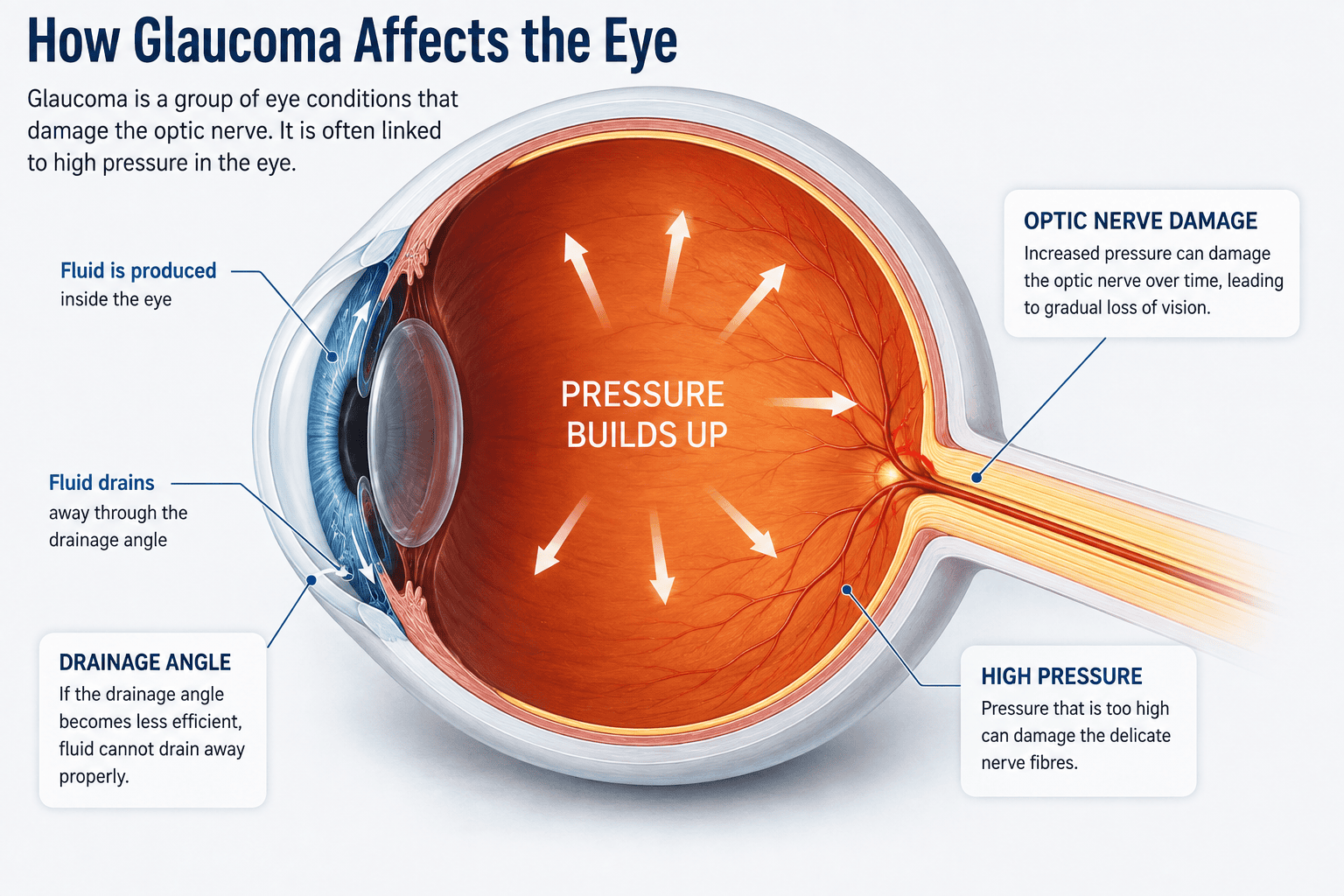
Glaucoma is the name given to a group of eye conditions that damage the optic nerve, which carries visual information from the eye to the brain.
The most common form, primary open-angle glaucoma, usually develops slowly and often causes no noticeable symptoms in its early stages.
Raised eye pressure is an important risk factor, but glaucoma is not simply another name for high eye pressure. Some people have raised pressure without optic-nerve damage, while others develop glaucoma despite having pressure measurements within the statistically normal range.
During an eye examination, the optometrist may assess the appearance of the optic nerve, eye pressure, the visual field and relevant risk factors such as age and family history.
OCT imaging can also help assess the retinal nerve-fibre layer and optic nerve and may be useful when comparing changes over time.
People with a close family history of glaucoma have a higher risk, and regular eye examinations are particularly important. In England, some people with glaucoma or a qualifying close family history may be entitled to a free NHS sight test; see our NHS eye examination eligibility guide.
Read our detailed guide to glaucoma, including symptoms, risk factors, tests and treatment.
Age-related macular degeneration
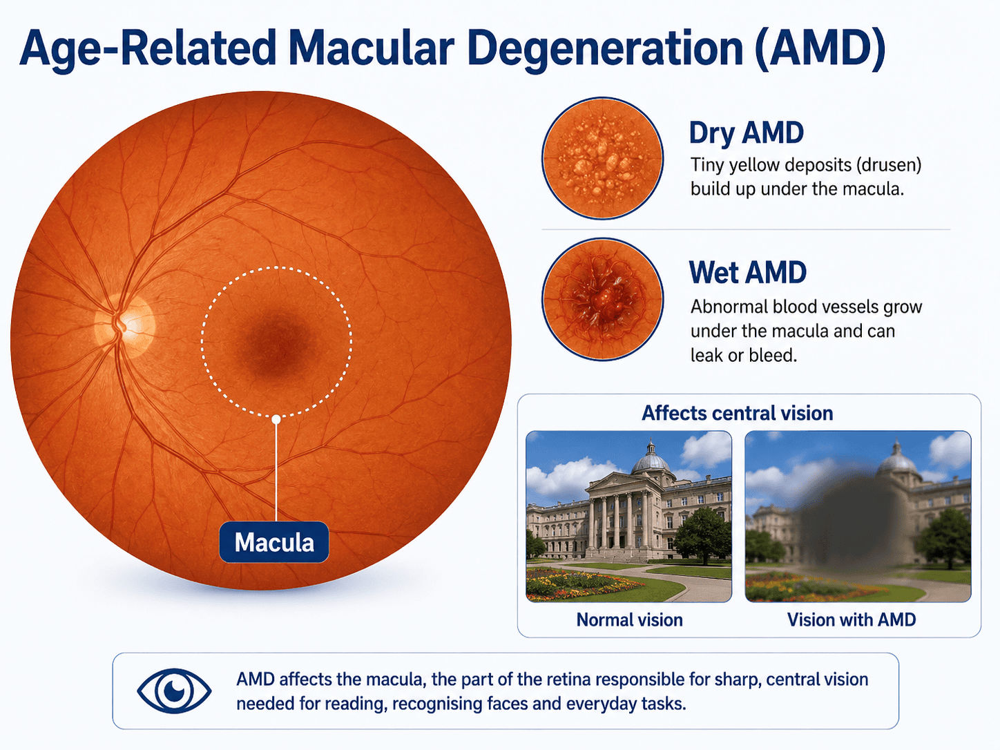
Age-related macular degeneration, usually called AMD, affects the macula — the small central part of the retina responsible for detailed central vision.
AMD becomes more common with increasing age and is generally divided into two main types: dry AMD and wet AMD.
Dry AMD
Dry AMD is the more common form and generally develops gradually. Changes can include deposits called drusen and changes to the retinal tissues around the macula.
People may notice increasing difficulty with reading, recognising faces, seeing fine detail or adapting between different lighting conditions. Some people with early dry AMD may notice very little change at all.
Wet AMD
Wet AMD is less common but can cause much more rapid deterioration in central vision. Abnormal blood vessels develop beneath or within the retina and can leak fluid or blood.
Symptoms may include straight lines appearing wavy, distortion in the centre of vision, a blurred or missing central area or a sudden deterioration in detailed vision.
Wet AMD needs prompt specialist assessment because treatment is generally more effective when started early.
OCT imaging is particularly useful when assessing the macula because it provides detailed cross-sectional images of the retinal layers and can show fluid or structural change.
Cataract
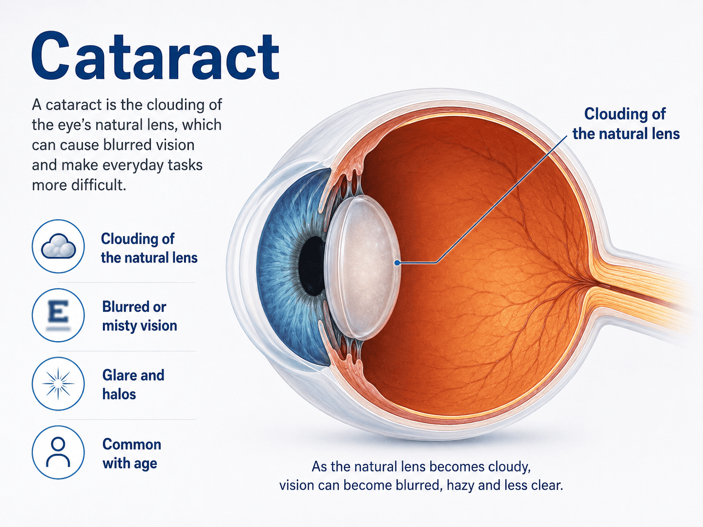
A cataract occurs when the normally clear natural lens inside the eye becomes cloudy.
Cataracts are extremely common as we get older and usually develop gradually.
Typical symptoms include blurred or misty vision, increased glare from headlights or bright sunlight, difficulty seeing at night, colours appearing less vivid and increasingly frequent changes to the spectacle prescription.
Cataracts are usually readily recognised during an eye examination. In their early stages, an updated glasses prescription may be enough to improve vision. As the cataract progresses, surgery may eventually be recommended.
Cataract surgery involves removing the cloudy natural lens and replacing it with a clear artificial lens. The timing of surgery depends largely on how much the cataract is affecting everyday life rather than simply on whether a cataract is present.
Read our detailed guide to cataract symptoms, causes, treatment and when to have an eye test.
Diabetes and the eyes
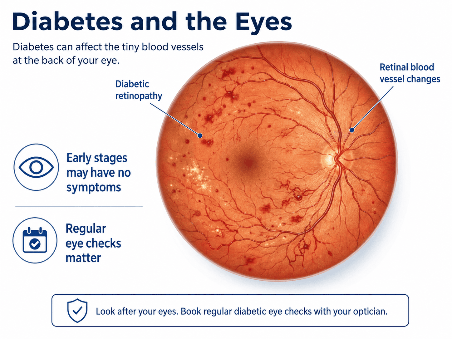
Diabetes can affect several structures within the eye, but one of the most important complications is diabetic retinopathy.
Persistently raised blood glucose can damage the small blood vessels within the retina. Early changes may cause no symptoms, while more advanced diabetic retinopathy can cause bleeding, swelling of the macula and loss of vision.
People with diabetes in England are offered dedicated NHS diabetic eye screening, which is separate from a routine sight test.
Regular eye examinations are still important because diabetes can also be associated with other eye changes, including fluctuations in vision, cataract and an increased risk of some other eye conditions.
An optometrist examining the retina may sometimes see changes associated with diabetes, but a routine eye examination should not be regarded as a substitute for attending diabetic eye-screening appointments.
Posterior vitreous detachment
The inside of the eye is filled with a transparent gel called the vitreous.
As we get older, the vitreous naturally becomes more liquid and can gradually separate from the retina. This is called a posterior vitreous detachment, or PVD.
PVD is very common, particularly later in life. Typical symptoms include new floaters, cobweb-like shapes, small dots or strands moving across the vision and flashes of light, particularly towards the edge of the vision.
Most PVDs do not cause lasting damage. However, occasionally the vitreous can pull hard enough on the retina to cause a retinal tear, so new flashes or a sudden increase in floaters should be assessed promptly.
Retinal tears and retinal detachment
A retinal tear can sometimes allow fluid to pass underneath the retina, causing it to separate from the back of the eye. This is called a retinal detachment.
Retinal detachment can threaten sight and needs urgent treatment.
Warning symptoms can include a sudden increase in floaters, repeated flashes of light, a dark curtain or shadow moving across the vision or sudden loss of part of the visual field.
Anyone experiencing these symptoms should seek urgent assessment rather than waiting for a routine eye examination.
Read our detailed guide to flashes and floaters and when to seek urgent help.
See our Urgent Eyecare information if you are experiencing sudden symptoms.
Epiretinal membrane
An epiretinal membrane is a thin layer of tissue that develops over the surface of the macula. It is more common with age and is sometimes discovered by chance during an eye examination.
If it contracts, it can slightly distort the underlying retina. Symptoms may include blurred central vision, straight lines appearing bent or wavy, difficulty with fine detail or differences between the vision in the two eyes.
OCT is particularly useful when assessing an epiretinal membrane because the scan shows the layers of the macula in detail. Many mild cases are simply monitored, while more significant cases may be referred for specialist assessment.
Macular hole
A macular hole is a small defect that forms in the centre of the macula.
It can cause blurred central vision, distortion, difficulty reading or a small missing area in the centre of vision.
Macular holes are more common with age and may develop as a result of traction from the vitreous. OCT imaging can usually show the structure of the macula clearly and plays an important role in assessment and monitoring.
Some macular holes require surgical treatment.
Dry eye disease
Dry eye disease occurs when the eyes do not produce enough tears, when tears evaporate too quickly, or when the tear film is unstable.
It is extremely common. Symptoms can include burning or stinging, gritty or sandy sensations, intermittent blurred vision, redness, watery eyes, discomfort when using screens and sensitivity to air conditioning or heating.
Many different factors can contribute, including age, medications, eyelid problems, contact lenses, screen use and conditions affecting the oil-producing glands in the eyelids.
Treatment depends on the underlying cause and may include lubricating drops, eyelid care and changes to the environment or daily routine.
Keratoconus
Keratoconus is a condition in which the cornea gradually becomes thinner and changes shape.
Instead of maintaining a regular curved surface, the cornea can become increasingly cone-shaped. This can cause increasing short sight, irregular astigmatism, distorted or ghosted vision, frequent prescription changes and difficulty achieving clear vision with ordinary spectacles.
Keratoconus often begins in teenagers or young adults. Specialist contact lenses can provide good vision for many people, and treatments such as corneal cross-linking may be recommended in progressive cases.
Early recognition is valuable because treatment decisions often depend on whether the condition is stable or changing.
Fuchs’ endothelial dystrophy
Fuchs’ endothelial dystrophy affects the innermost layer of the cornea. The endothelial cells help keep the cornea clear by pumping excess fluid out of it.
If too many of these cells are lost, the cornea can begin to swell. Symptoms may include misty or blurred vision, vision that is worse on waking, glare and halos around lights.
The condition usually develops slowly and is often detected during an eye examination before it causes significant difficulty. More advanced cases may require specialist treatment.
Uveitis
Uveitis is inflammation inside the eye. It can affect different parts of the eye and has a wide range of possible causes.
Symptoms can include eye pain, redness, sensitivity to light, blurred vision and floaters.
Unlike many chronic eye conditions, uveitis can develop relatively quickly. A painful, red or light-sensitive eye should therefore be assessed rather than waiting for the next routine examination.
Why regular eye examinations matter
A sight test is not simply a check to see whether you need new glasses.
During an eye examination, your optometrist can assess many aspects of eye health and compare current findings with previous examinations.
This is particularly valuable because changes over time can sometimes be more informative than a single measurement.
Depending on your age, symptoms, family history and examination findings, this may include examination of the retina and optic nerve, eye-pressure measurement, visual-field testing, assessment of the front of the eye, pupil reactions and OCT imaging where appropriate.
Find out more about Eye Examinations at Actons Opticians.
How OCT can help
Optical Coherence Tomography, or OCT, produces detailed cross-sectional images of the retina and optic nerve.
It can provide useful additional information when assessing or monitoring conditions affecting structures such as the macula, retinal nerve-fibre layer, optic nerve and vitreoretinal interface.
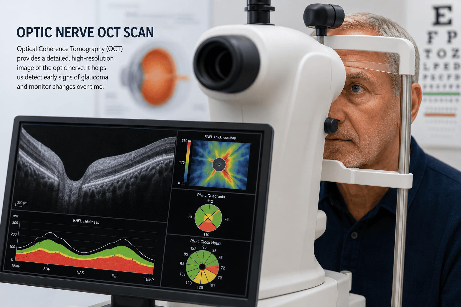
At Actons Opticians we use a Heidelberg Engineering Spectralis® OCT.
An important advantage of building up OCT records over time is the ability to compare the same structures at different visits. This can help distinguish something that is naturally different in an individual eye from something that may actually be changing.
OCT does not replace a full eye examination and it cannot diagnose every eye condition by itself. It is one part of the overall clinical assessment.
Read more about OCT Eye Scans in Worcester
Why comparing OCT eye scans over time can be useful
Frequently asked questions about eye conditions
| Question | Answer |
|---|---|
| Can an eye test detect eye disease before I notice symptoms? | Sometimes, yes. A routine eye examination can reveal signs or changes affecting structures such as the retina, optic nerve, cornea and natural lens before you are aware of a problem yourself. This is one reason regular eye examinations are important, particularly if you have risk factors such as age, diabetes or a family history of eye disease. If something looks unusual, your optometrist can advise whether it should be monitored, investigated further or referred for specialist assessment. |
| Does OCT diagnose eye conditions? | OCT is a very useful imaging test, but it does not diagnose every eye condition on its own. It provides detailed cross-sectional images of the retina and optic nerve and can help an optometrist assess or monitor conditions such as glaucoma, macular disease, epiretinal membranes and macular holes. OCT findings are interpreted alongside the rest of the eye examination, which may include symptoms, vision, eye pressure, visual fields and examination of the eye itself. |
| How often should I have an eye examination? | For many adults, a routine eye examination every two years is a common interval, but some people need to be seen more often. Your optometrist may recommend a shorter interval if you have an eye condition, significant risk factors, certain medical conditions or findings that need monitoring. If you develop new symptoms, do not wait until your next routine appointment. |
| Which eye symptoms need urgent attention? | Sudden loss of vision, a new curtain or shadow across the sight, a sudden shower of floaters, persistent flashes of light, severe eye pain, a very red painful eye or sudden marked distortion of central vision can all require prompt assessment. If symptoms are sudden or severe, follow the advice on our Urgent Eyecare page rather than waiting for a routine sight test. |
| Does having a family history increase my risk of eye disease? | For some conditions, yes. Glaucoma is a good example: having a close relative with glaucoma increases your risk. Family history can also be relevant in other conditions such as some corneal and retinal disorders. Tell your optometrist about important eye conditions that run in your family so this can be taken into account during your examination. |
| Can diabetes affect my eyes even if my vision seems normal? | Yes. Diabetic retinopathy can develop without obvious visual symptoms in its early stages. This is why people with diabetes should attend their NHS diabetic eye-screening appointments as recommended. Routine eye examinations remain important too, but they are not a substitute for the dedicated diabetic eye-screening programme. |
| Can cataracts, glaucoma or AMD be prevented? | There is no guaranteed way to prevent all cases of these conditions. Age and genetics play an important part in many eye diseases. The most useful strategy is to reduce avoidable risk where possible, look after general health and have regular eye examinations so that significant changes can be identified early. For conditions such as glaucoma, early detection and treatment can substantially reduce the risk of avoidable sight loss. |
When symptoms need urgent attention
Some eye conditions develop slowly. Others need much quicker assessment.
Seek urgent help if you experience symptoms such as sudden loss of vision, a curtain or dark shadow across your sight, a sudden shower of new floaters, persistent flashing lights, severe eye pain, a very red painful eye or sudden marked distortion or loss of central vision.
If you are experiencing sudden or worrying symptoms, see our Urgent Eyecare in Worcester page rather than waiting for a routine appointment.
Concerned about your eye health?
Many eye conditions are easier to manage when changes are identified early.
Even when your vision seems unchanged, a comprehensive eye examination allows us to assess much more than your spectacle prescription.
At Actons Opticians in Worcester we take time to assess your vision, eye health and any individual risk factors. Where appropriate, additional testing such as visual-field assessment or Spectralis OCT imaging can provide further information.
If you have a family history of an eye condition, have noticed a change in your vision, or are simply due your next examination, you can arrange an appointment with us.
Would you like to arrange an eye examination?
Contact Actons Opticians to arrange an eye examination or to discuss any concerns about your eye health.