Glaucoma is one of the most important eye conditions to detect early because it can gradually damage the optic nerve and cause permanent loss of vision, often without producing obvious symptoms at first.
Many people who develop glaucoma feel that their eyesight is perfectly normal when the condition is first identified. In fact, glaucoma is commonly discovered during a routine eye examination rather than because someone has noticed a problem themselves.
The good news is that modern treatments are usually very effective at reducing the risk of further damage. The vision already lost through glaucoma cannot normally be restored, but identifying the condition early and controlling it can often preserve useful sight for life.
This guide explains what glaucoma is, why it develops, who is at greater risk, the different types, how optometrists look for the early signs, and what treatment and lifestyle measures may help.
If you would like a broader overview of other conditions that can affect the eyes, including AMD, cataract and diabetic eye disease, see our Common Eye Conditions guide.
What is glaucoma?
Glaucoma is the name given to a group of eye diseases that damage the optic nerve.
The optic nerve is the cable-like structure that carries visual information from the retina at the back of the eye to the brain. For us to see normally, the optic nerve needs to remain healthy.
In glaucoma, some of the nerve fibres gradually become damaged and die. As this progresses, areas of the visual field can be lost.
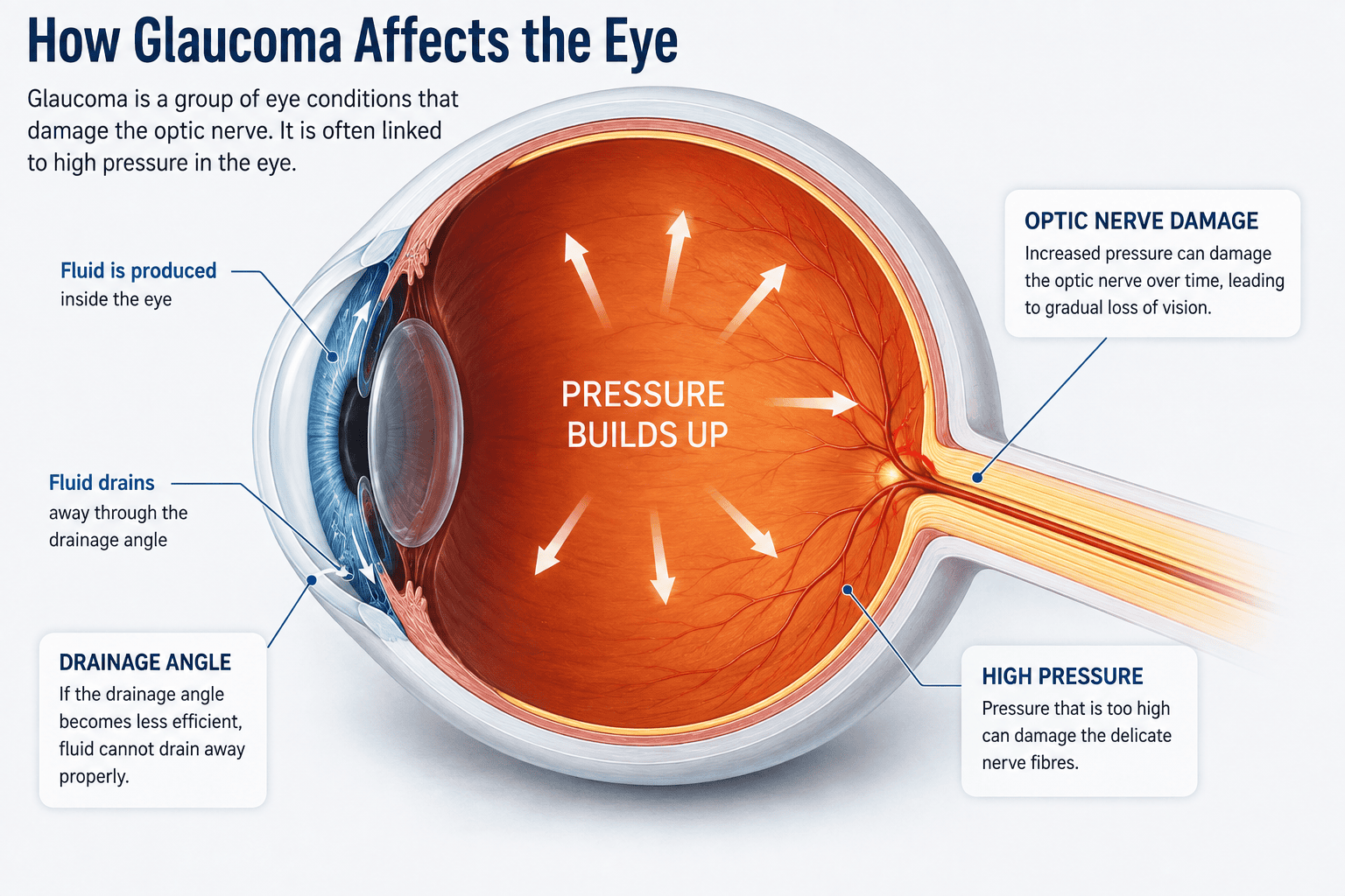
A common misconception is that glaucoma simply means high pressure in the eye. Raised eye pressure is an important risk factor, but glaucoma is actually defined by damage to the optic nerve.
Some people have high eye pressure but never develop glaucoma. This is called ocular hypertension.
Conversely, some people develop glaucoma even though their measured eye pressure is within what would traditionally be considered the normal range. This is often called normal-tension glaucoma.
What is eye pressure?
The front part of the eye contains a clear fluid called aqueous humour.
This fluid is continuously produced inside the eye and normally drains away through microscopic drainage channels around the edge of the iris and cornea.
If the fluid does not drain efficiently enough, pressure can build inside the eye. This pressure is known as intraocular pressure, usually shortened to IOP.
Eye pressure is measured in millimetres of mercury, or mmHg.
There is no single pressure at which everyone develops glaucoma. Some optic nerves tolerate relatively high pressures without damage, while others may be more vulnerable.
This is why an eye-pressure measurement alone cannot diagnose or exclude glaucoma.
What happens to your vision if you develop glaucoma?
The common forms of glaucoma usually affect the peripheral visual field before central vision.
This means the condition can progress significantly without someone noticing that anything is wrong.
The brain is also remarkably good at filling in missing visual information, and one eye may compensate for changes in the other.
As glaucoma progresses, areas of peripheral vision can become permanently damaged. Eventually, if the condition is severe and untreated, the remaining field of vision can become increasingly restricted.
Advanced glaucoma can ultimately affect central vision as well.
The important point is that glaucoma damage is irreversible. Treatment is therefore designed to protect the vision that remains rather than restore nerve tissue that has already been lost.
This is why early detection is so valuable.
What causes glaucoma?
For many people, there is no single identifiable cause.
The commonest form, primary open-angle glaucoma, usually develops gradually as the eye’s drainage system becomes less efficient with age.
Pressure within the eye may increase, and over time this can contribute to damage to vulnerable optic-nerve fibres.
However, the relationship between pressure and glaucoma is complex. Blood supply to the optic nerve, inherited characteristics of the nerve, age and other biological factors may all influence how vulnerable an individual optic nerve is.
There are also forms of glaucoma caused by another eye condition, medication, injury or abnormal development of the eye.
What are the different types of glaucoma?
Primary open-angle glaucoma
Primary open-angle glaucoma (POAG) is the most common form in the UK.
The drainage angle of the eye remains physically open, but fluid does not drain away efficiently enough through the trabecular meshwork.
The condition normally develops slowly over many years.
There is usually no pain, redness or noticeable change in eyesight in the early stages. That is why regular eye examinations are important.
Normal-tension glaucoma
Normal-tension glaucoma causes characteristic optic-nerve damage and visual-field loss even though measured eye pressure lies within the statistically normal range.
The exact mechanism is not fully understood. Some optic nerves may simply be more vulnerable to a given level of pressure, and the blood supply to the optic nerve may also be important.
Treatment still usually involves lowering eye pressure because reducing pressure has been shown to lower the risk of further damage.
Ocular hypertension
Ocular hypertension is not glaucoma, but it is closely related.
It means that eye pressure is consistently higher than expected without detectable damage to the optic nerve or visual field.
Some people with ocular hypertension never develop glaucoma, while others do.
Risk assessment considers factors such as the actual eye pressure, corneal thickness, age, family history and life expectancy.
Primary angle-closure glaucoma
In angle-closure glaucoma, the drainage angle between the iris and cornea becomes narrowed or closed, preventing fluid from leaving the eye normally.
This can happen gradually, but occasionally the drainage angle closes suddenly.
A sudden attack of acute angle closure can cause a rapid and potentially dangerous rise in eye pressure.
Symptoms can include:
- severe eye pain
- a very red eye
- blurred vision
- coloured halos around lights
- headache
- nausea or vomiting
Acute angle closure is an eye emergency and requires immediate treatment.
Secondary glaucoma
Secondary glaucoma occurs because of another condition or external factor.
Possible causes include:
- inflammation inside the eye
- eye injury
- certain retinal conditions
- abnormal blood vessels inside the eye
- previous eye surgery
- pigment or material blocking the eye’s drainage system
- some medicines, particularly corticosteroids in susceptible people
Steroid medication can increase eye pressure in some individuals. This can occur with steroid eye drops but can also happen with other forms of steroid treatment. Anyone using long-term steroids should make sure their eyecare professional knows about them.
Developmental and childhood glaucoma
Glaucoma can very occasionally affect babies and children because the eye’s drainage structures have not developed normally.
This is very different from the common age-related form of glaucoma and usually requires specialist ophthalmological care.
Who is most at risk of glaucoma?
Anyone can develop glaucoma, but several factors significantly increase the likelihood.
Age
Age is one of the strongest risk factors.
Primary open-angle glaucoma is relatively uncommon in younger adults but becomes progressively more common with each decade of life.
Family history
Glaucoma often runs in families.
Having a parent, brother, sister or child with glaucoma increases your risk considerably.
This is why patients diagnosed with glaucoma are often encouraged to tell their close relatives.
In England, people aged 40 or over with a parent, sibling or child who has glaucoma may also qualify for an NHS-funded sight test.
Ethnic background
Risk varies between populations.
People of African or African-Caribbean ancestry have a substantially higher risk of developing primary open-angle glaucoma and may develop it at a younger age.
People of East Asian and some South Asian backgrounds have a higher risk of certain forms of angle-closure glaucoma.
High eye pressure
Raised intraocular pressure is the most important modifiable risk factor for glaucoma.
However, high pressure does not automatically mean glaucoma and a normal pressure does not completely rule it out.
Short sight
People who are significantly short-sighted, particularly those with higher levels of myopia, have an increased risk of open-angle glaucoma.
Highly myopic optic nerves can also be more difficult to assess because their appearance may differ from average.
Long sight
Long-sighted eyes tend to be smaller and can have shallower anterior chambers.
This can increase the likelihood of a narrow drainage angle and therefore angle-closure glaucoma in susceptible individuals.
Thin corneas
People with relatively thin central corneas have a greater risk of developing glaucoma in some circumstances.
Corneal thickness also affects the interpretation of eye-pressure measurements, which is one reason hospital glaucoma assessments often include a corneal-thickness measurement.
Diabetes and blood pressure
Diabetes is associated with a higher risk of glaucoma in some studies.
Blood-pressure relationships are more complicated. Very high blood pressure may be associated with raised eye pressure, while unusually low blood pressure may reduce blood flow to the optic nerve in susceptible people.
Steroid use
Some people are particularly sensitive to corticosteroids and experience a significant rise in eye pressure when using them.
Risk may be greater with prolonged treatment and in people who already have glaucoma or other risk factors.
Never stop prescribed steroid treatment without medical advice, but make sure your optometrist and ophthalmologist know what medication you use.
What symptoms does glaucoma cause?
This depends on the type.
Chronic open-angle glaucoma
The most common form usually causes no symptoms in its early stages.
There is normally:
- no pain
- no redness
- no sudden blur
- no obvious awareness of peripheral vision being lost
This is what makes glaucoma potentially dangerous.
Many people only discover that they have it because an optometrist notices suspicious changes during a routine eye examination.
Acute angle closure
Acute angle closure is very different and can cause dramatic symptoms such as severe pain, redness, blurred vision, halos, headache and nausea.
Anyone developing these symptoms suddenly should seek immediate emergency medical attention. NHS guidance advises calling 999 or going to A&E if glaucoma symptoms develop suddenly, as acute angle closure can threaten sight and may require urgent treatment.
How can an optometrist detect glaucoma early?
An eye examination is about much more than checking whether your spectacle prescription has changed.
Optometrists routinely look for signs that may indicate glaucoma, especially in patients who have recognised risk factors.
There is no single glaucoma test. Instead, several pieces of information are considered together.
Examining the optic nerve
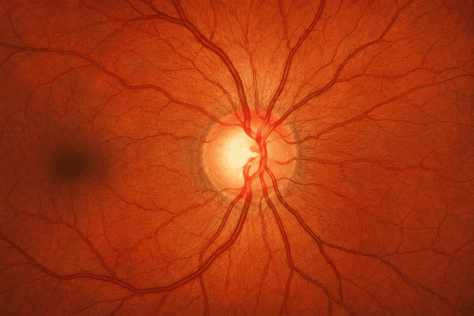
The optometrist examines the optic nerve head, also called the optic disc.
Glaucoma can produce characteristic changes in its shape and appearance, sometimes described as increased cupping.
However, optic nerves naturally vary enormously between individuals. A large cup does not automatically mean glaucoma.
This is why comparison with previous records and images can be particularly useful.
Measuring eye pressure
Eye pressure can be measured in several ways.
It is useful information but should never be considered in isolation because people with normal pressures can develop glaucoma and some people with relatively high pressures remain healthy.
Visual-field testing
A visual-field test assesses how much of the surrounding visual world each eye can detect while you look straight ahead.
Glaucoma can produce characteristic areas of missing or reduced sensitivity in the visual field.
Repeating visual-field tests over time can also help determine whether an eye is stable or progressing.
OCT imaging
Optical Coherence Tomography, or OCT, allows extremely detailed imaging of structures around the optic nerve and retina.
In glaucoma assessment, OCT can measure areas such as the retinal nerve-fibre layer, which contains the nerve fibres travelling towards the optic nerve.
Changes in these measurements can sometimes help identify structural damage or monitor progression.
At Actons Opticians we use a Heidelberg Engineering Spectralis® OCT, which allows accurately aligned follow-up scans to be compared over time.
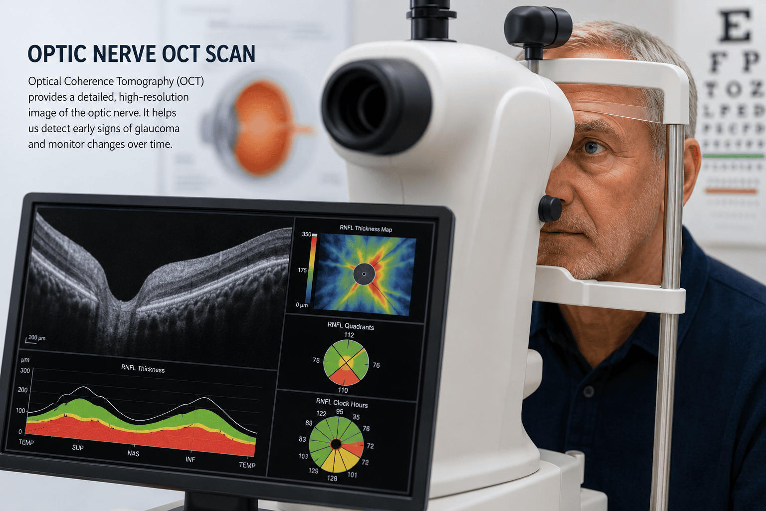
However, OCT does not replace the rest of the glaucoma assessment. Structural OCT information and functional visual-field testing are complementary rather than interchangeable.
You can read more about our OCT Eye Scans in Worcester and why comparing OCT scans over time can be useful.
Assessing the drainage angle
If someone is at risk of glaucoma, the optometrist may also assess the depth and configuration of the front chamber of the eye.
This can help identify people whose drainage angle may be unusually narrow and therefore at risk of angle closure.
What happens if an optometrist suspects glaucoma?
An optometrist does not normally make a definitive diagnosis of glaucoma solely from a routine eye examination.
If the combination of eye pressure, optic-nerve appearance, visual field, OCT or other findings raises concern, further assessment may be recommended.
Depending on local arrangements, this may involve a specialist optometrist, glaucoma referral-refinement service or hospital eye department.
Providing previous measurements and images can be particularly useful when deciding whether something is genuinely changing.
How is glaucoma treated?
The central aim of glaucoma treatment is to lower eye pressure sufficiently to reduce the risk of further optic-nerve damage.
The appropriate treatment depends on the type of glaucoma, the pressure level, how much damage is already present, the patient’s age and other individual factors.
Eye drops
Pressure-lowering eye drops have been used successfully for many years.
Different types work either by reducing the amount of fluid produced inside the eye or by helping it drain more efficiently.
Some patients need one type of drop, while others need a combination.
Using the drops regularly is extremely important. Glaucoma often causes no symptoms, so it can be tempting to forget treatment when the eyes feel completely normal.
Selective laser trabeculoplasty
Selective laser trabeculoplasty, usually shortened to SLT, is now an important treatment for open-angle glaucoma and ocular hypertension.
The laser is applied to the eye’s drainage tissue to improve the outflow of fluid and reduce eye pressure.
For suitable patients it can reduce or delay the need for daily eye drops.
Surgery
If eye drops and laser treatment do not control the pressure adequately, glaucoma surgery may be recommended.
Traditional glaucoma surgery, such as a trabeculectomy, creates a new pathway for fluid to leave the eye.
There are also drainage implants and a growing range of less invasive glaucoma procedures.
The exact treatment is selected by the ophthalmologist according to the type and severity of disease.
Treatment of angle closure
Angle-closure glaucoma is treated differently.
Laser treatment may be used to create a tiny opening in the iris so that fluid can circulate more freely and the drainage angle is less likely to become blocked.
Acute attacks require urgent treatment to lower pressure quickly and protect the optic nerve.
Can glaucoma be cured?
At present, damage already caused by glaucoma cannot normally be reversed.
For that reason glaucoma is usually regarded as a long-term condition that needs monitoring rather than something that is cured and forgotten.
However, treatment can be extremely effective.
The majority of people whose glaucoma is detected and treated appropriately do not become blind.
The prognosis is generally best when glaucoma is discovered before significant visual-field loss has occurred and when treatment and follow-up are maintained.
Can glaucoma be prevented?
There is no guaranteed way to prevent the common inherited and age-related forms of glaucoma.
You cannot change your age, genetics or ethnic background.
The most effective strategy for preventing avoidable sight loss is therefore early detection and appropriate treatment.
Regular eye examinations are particularly important if you have a family history or another recognised risk factor.
Can lifestyle reduce glaucoma risk?
This is an area where it is important not to overstate the evidence.
A healthy lifestyle is good for overall cardiovascular and eye health, but there is currently no diet, supplement or exercise programme proven to prevent glaucoma.
Lifestyle should complement proper eye examinations and prescribed treatment, not replace them.
Exercise
Regular moderate exercise is generally beneficial and some studies suggest that aerobic activity can temporarily lower eye pressure.
Walking, cycling and similar activity are therefore sensible parts of a healthy lifestyle.
If you already have glaucoma, very heavy resistance exercise involving prolonged breath-holding can temporarily raise eye pressure. Certain inverted yoga positions, where the head remains substantially below the heart, can also cause a temporary pressure increase.
For most people these short-term changes are unlikely to be important, but someone with advanced glaucoma should discuss unusually strenuous or inverted exercise with their eye specialist.
Diet
There is no particular “glaucoma diet”.
Eating a balanced diet rich in vegetables, fruit, whole grains, fish and other nutritious foods is worthwhile for general health, but no specific food has been proven to prevent glaucoma.
Supplements
There is currently insufficient evidence to recommend nutritional supplements specifically to prevent or treat glaucoma.
Research is ongoing into several nutrients and potential neuroprotective treatments, but none should replace proven pressure-lowering treatment.
Caffeine
Large amounts of caffeine can temporarily raise eye pressure in some people.
There is no need for most people to avoid tea or coffee completely, but moderation is sensible, particularly for someone known to have glaucoma or high eye pressure.
Smoking
Smoking has many established harmful effects on general health and other aspects of eye health.
Evidence linking smoking specifically to glaucoma progression is less straightforward, but avoiding smoking remains one of the best general health measures anyone can take.
Blood pressure and general health
Keeping cardiovascular risk factors such as blood pressure and diabetes appropriately managed is sensible for both general and eye health.
However, patients should not attempt to deliberately lower their blood pressure simply because they have glaucoma. Excessively low blood pressure may be undesirable for optic-nerve perfusion in some people.
Medication changes should always be discussed with the relevant doctor.
The most important glaucoma risk-reduction measures
- Have regular eye examinations, even if your sight seems completely normal.
- Tell your optometrist about glaucoma in your family.
- Mention all medications, particularly steroid treatment.
- Attend follow-up appointments if you have ocular hypertension or suspicious optic-nerve findings.
- Use prescribed glaucoma treatment consistently.
- Encourage close relatives to have their eyes examined if you are diagnosed with glaucoma.
- Maintain a generally healthy lifestyle, while recognising that lifestyle measures cannot substitute for clinical monitoring and treatment.
Do family members of someone with glaucoma need testing?
Yes, regular eye examinations are especially important for close relatives.
A parent, brother, sister or child of somebody with glaucoma has a significantly higher risk than the population as a whole.
Because the common forms are usually symptomless at first, waiting until vision changes is not a good strategy.
In England, people aged 40 or over who have a parent, sibling or child diagnosed with glaucoma can qualify for a free NHS sight test.
You can read more on our NHS Eye Examination Eligibility page.
When should I arrange an eye examination?
Do not wait for symptoms if you are due a routine examination.
It is particularly worth making sure your examinations are up to date if:
- glaucoma runs in your family
- you are over 40
- you have high eye pressure
- you are significantly short-sighted
- you have diabetes
- you use long-term steroid medication
- you have previously been told that your optic nerves look unusual
- you have ocular hypertension
- you belong to an ethnic group known to have a higher glaucoma risk
If you suddenly develop severe eye pain, a red eye, blurred vision, coloured halos around lights, headache or nausea, seek urgent medical advice because acute angle closure is an emergency.
Glaucoma FAQs
| Question | Answer |
|---|---|
| Can you have glaucoma without high eye pressure? | Yes. Some people develop characteristic glaucoma damage even though their measured eye pressure is within the statistically normal range. This is often called normal-tension glaucoma. It is one reason why eye pressure should never be considered on its own. |
| Can glaucoma be detected during a routine eye test? | A routine eye examination can identify signs that may suggest glaucoma, such as changes in the optic nerve, raised eye pressure or characteristic visual-field defects. OCT imaging may provide additional structural information. If the findings are concerning, further assessment or referral may be recommended. |
| Can glaucoma be cured? | Glaucoma cannot usually be cured in the sense of reversing damage that has already occurred. However, treatment can lower eye pressure and significantly reduce the risk of further optic-nerve damage. Many people with glaucoma retain useful vision throughout life when the condition is detected and managed appropriately. |
| Does glaucoma always cause symptoms? | No. The most common form, primary open-angle glaucoma, usually develops slowly and often causes no noticeable symptoms in its early stages. Acute angle-closure glaucoma is different and can cause sudden severe pain, redness, blurred vision, halos, headache or nausea. |
| Is glaucoma hereditary? | Family history is an important risk factor. Having a parent, brother, sister or child with glaucoma increases your own risk, so close relatives should make sure their eye examinations are up to date. |
| How often should someone at higher risk of glaucoma have their eyes examined? | There is no single interval that is right for everyone. The appropriate frequency depends on factors such as age, family history, eye pressure, optic-nerve appearance and any previous findings. Your optometrist can advise how often you should be reviewed. In England, people aged 40 or over with a parent, sibling or child who has glaucoma may qualify for an NHS-funded sight test. |
Looking after your eyes over the long term
One of the reasons glaucoma deserves so much attention is that early disease often gives very little warning.
A person can read the smallest letters on an eye chart and still have early glaucoma-related damage elsewhere in their visual field.
That is why an eye examination looks beyond your spectacle prescription.
At Actons Opticians in Worcester, we assess eye health as part of a full eye examination and can use additional tests such as eye-pressure measurement, visual-field assessment and Spectralis OCT imaging when clinically appropriate.
Building up accurate records over several examinations can also be valuable. Comparing optic-nerve appearance, visual fields and OCT scans over time can help distinguish between a feature that is simply normal for your eyes and one that is genuinely changing.
If you have a family history of glaucoma, have previously been told that your eye pressure is raised, or simply want to make sure your eye examination is up to date, please mention any concerns when you arrange your appointment.
Find out more about Eye Examinations at Actons Opticians
Read about OCT Eye Scans in Worcester
Check NHS eye examination eligibility
Would you like to arrange an eye examination?
Contact Actons Opticians if you are concerned about glaucoma, have a family history of the condition or would like to arrange an eye examination.
Sources and further reading
- NHS: Glaucoma
- NICE: Glaucoma – diagnosis and management (NG81)
- College of Optometrists: Examining patients at risk from glaucoma
- Glaucoma UK: What is glaucoma?
Article reviewed against current UK guidance in August 2026.
Back to Eyecare for more related services and eye health advice.